12-lead Electrocardiogram (EKG)
 Your physician may order an EKG for the following reasons:
- identify the cause of an arrhythmia
- pursue evidence of a high-risk arrhythmia syndrome, such as Long-QT and Brugada Syndrome
- investigate a history of syncope or palpitations
- monitor a medication effect
- determine if there is evidence of a myocardial infarction, or “heart attack”
- investigate any evidence of coronary artery blockages
- determine normal pacemaker or ICD functioning
A standard surface EKG is recorded using electrodes, or sticky patches, placed on the body surface, typically over the chest and the extremities. These electrode wires are connected to the EKG machine with recordings from 12 different locations on the surface of the body, the so-called12-lead EKG. The resultant electrocardiogram represents a mesh of cardiac electrical activity from the atrium and ventricles, dependent on the direction and magnitude of the electrical depolarization as it spreads throughout the heart. It is recorded as an output of waveforms that your physician can print onto paper or record on the monitor.
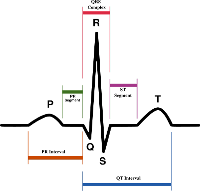
Figure 1. The components of an EKG recording during a single cardiac cycle (single heart beat) is shown above. The p-wave represents atrial activation, the QRS represents ventricular contraction, and the t-wave represents ventricular electrical recovery.
The waveforms depicted in an EKG can be divided into time segments measuring particular phases of the cardiac cycle or contraction. The continuous repetitive unit of the cardiac cycle is the “P-QRS-T” waveform. The p-wave represents the atrial depolarization, or contraction phase, when the sinus rhythm beat electrically captures the left and right atria. The QRS represents the depolarization or contraction of the lower ventricles. The duration of the QRS is indicative of ventricular disease in general, with a wider QRS representative of slowed ventricular conduction. The PR-interval is a representation of the time for atrial contraction to reach the ventricular muscle tissue, as it traverses the AV node. The QT-interval represents both ventricular activity and recovery, with the t-wave being specifically the recovery phase. Because the heart is continuously beating, this creates the consecutive strings of P-QRS-T waves as depicted below. An arrhythmia will alter the activation of the heart and the resultant EKG indices (including the sequence and directionality of activation). Information regarding the origin of your cardiac arrhythmia (which cardiac chamber), the presence of baseline electrical conduction system disease, signs of damage to the heart from a previous “heart attack”, the presence of medications, and normal pacemaker or ICD function can be gleaned from your 12-lead EKG.
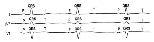
Figure 2. Continuous EKG recording from three different body surface leads (leads I, aVF, and V1).
Each of the 12 EKG leads represent a different direction of cardiac activation in 3-D space. The standard EKG leads are denoted as lead I, II, III, aVF, aVR, aVL, V1, V2, V3, V4, V5, V6. Leads I, II, III, aVR, aVL, aVF are denoted the limb leads while the V1, V2, V3, V4, V5, and V6 are precordial leads.
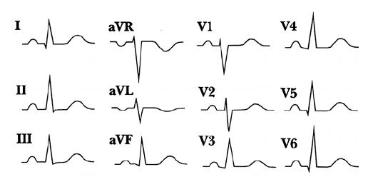
Figure 3. Basic appearance of the normal EKG recordings from the 12 surface leads producing the standard 12-lead EKG.
The surface recording electrodes of the EKG machine are placed in the same standard position each time, allowing for comparison of an individual’s EKG over time. This can help your physician to determine if you have had a “heart attack”, or a new arrhythmia. The 12-lead EKG provides more information on the diagnosis of your cardiac arrhythmia than an outpatient Holter or Event monitor, as it represents information recorded from a larger surface area surrounding the heart. However, cardiac arrhythmias tend to be transient and episodic, making capture of the 12-lead EKG impractical during these episodes.


 Silver Spring Office
  DC Office (at Providence Hospital)
  Hagerstown Office